3531376
89 y/o man, heart failure
1414720
82 y/o man, FUO
2010年5月2日 星期日
2010年3月13日 星期六
physical examination of cardiovascular system
abdomen:
pulsatile, expansible abdominal, mass: aortic aneurysm
large, tender liver: heart failure, constrictive pericarditis
systolic hepatic pulsation: TR
palpable spleen: severe heart failure, infective endocarditis
ascites: heart failure, constrictive heart failure (especially when ascites is out of proportion of limbs edema)
extremities:
ankle-brachial index (ABI): consider to be abnormal when <0.9, critical stenosis when < 0.3
thrombophlebitis: pain in the calf or thigh, with edema
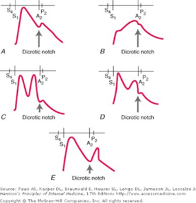
arterial pressure pulse:
pulsus parvus: small weak pulse, common condition with decreased cardiac output
pulsus tardus: delayed systolic pulse, severe AS
pulsus bisferiens: two systolic peak, AR, or hypertrophic cardiomyopathy
pulsus alternans: regular rhythm with regular alternation of the pressure pulse amplitude, severe impairment of
LV function
pulsus paradoxus: systolic pressure decreased> 10 mmHg when inspiration, cardiac temponade, severe
lung disease, severe vena cava obstruction
a: atrial contraction
c: TV bulging into RA when RV isovolumetric systole
x: atrial relaxation when RV contraction
v: right atrial volume increased when RV systole and TV close
y: RV pressure decline and TV open
abdominal-jugular reflex: increased upper level of venous pulsation when abdominal compression (>10 seconds), indicate heart failure or TR
Kussmaul sign: increased rather than decreased CVP level during inspiration, indicate severe right-heart
failure
Precordial palpation:
normal LV apex impulse: mid-clavicular line, 4th or 5th intercostal space
LV hypertrophy: lateral and downward displacement of LV apex impulse
RV hypertrophy: sustained systolic lift at lower left sternal border
MR: left parasternal lift due to RV anterior displacement (compressed by enlarged LA)
visible or palpable pulmonary flow over the left 2nd intercostal space: pulmonary hypertension
Cor pulmonale
definition:
pulmonary heart disease, excluding congenital heart disease, or right heart disease induced by left heart
etiology:
any pulmonary vascular or parenchymal disease can lead to cor pulmonale
50% cases were due to COPD
pathophysiology:
RV failure due to elevated pulmonary vascular pressure
acute: RV dilation without hypertrophy
chronic: RV dilation and hypertrophy
symptoms and signs
dyspnea, orthopnea, limbs edema, ascites
holosystolic murmur (TR murmur), increased with inspiration (Carvallo's sign)
Prominent V waive
diagnosis
1. exclude LV failure induced RV failure
2. EKG: P pulmonale, RV hypertrophy, right axis deviation
3. EKG: engorged pulmonary artery
treatment:
1. treat underlying pulmonary diseases
2. diuretics
3. digoxin: unclear role, should be administered with low dose
2010年3月3日 星期三
heart failure
definition
abnormal cardiac structure or function, which leads to signs (edema, rale) and symptoms (fatigue, dyspnea )
etiology:
75% due to CAD and HTN in developed country
pathogenesis
1. index events
2. compensatory mechanism
a. RAA system
b. adrenergic nerve system
c. increased myocardial contratility
basic mechanism
1. systolic dysfunction
2. diastolic dysfunction
3. LV remodeling
Clinical menifestation
1. fatigue and dyspnea
2. orthopnea and nocturnal cough
3. Paroxysmal nocturnal dyspnea
4. Cheyne-Stoke respiration
5. Acute lung edema
6. Anorexia, nausea
7. Nocturia
Physical examination
1. reduced BP and pulse pressure
2. cool extremities
3. Jugular vein engorgement
4. rales and crackles (may be absent in chronic HF due to increased lymphatic drainage)
5. pleural effusion (drain to systemic and pulmonary vein, means bi-ventricular failure)
6. displaced the point of maximal impulse (PMI), usually displaced below the fifth intercostal space and/or lateral to the midclavicular line
7. S3, S4, MR and TR murmur in advaned cases
8. hepatomegaly, ascites
9. peripheral edema
Diagnosis
1. Clinical symptoms/ signs
2. routine lab
3. ECG
4. CXR
5. cardiac echo/ MRI
6. cardiac enzyme
Treatment
1. stage: A: high risk patients without structural abnormality or symptoms
B: structural abnormality without symptoms
C: structural heart disease with symptoms
D: refractory heart failure
2. factors may precipitate acute decompensation in chronic heart failure patient
a. dietary
b. discontinue treatment
c. MI
d. arrhythmia
e. infection
f. anemia
g. medication:
NSAID, CCB, Beta-blocker, Class I anti-arrhythmic drugs
h. alcohol
i. pregnancy
j. worsing hypertension
k: acute valvular disease
3. Diet
sodium: 2~3 g/ day
fluid restriction (< 2 L/ day) is not generally necessary
4. Diuretics
5. ACEI
6. Beta-blocker
7. Aldactone
8. Hydralazine/ Ismo-20
9. Digoxin
Acute heart failure
Theraputic goal
1. stablize hymodynamic
2. treat reversible factors
3. reestablish an effective outpatient medical regimen
LV filling pressure: elevated: wet normal: dry
Cardiac output: decreased: cold normal: warm
Pharmacological management
1. Diuretics
2. Vasodilators
3. Inotropic agents
Mechanical management:
1. IABP
2. ECMO
3. LV assist device
4. heart transplant
2010年2月22日 星期一
2010年2月21日 星期日
diabetes ketoacidosis (DKA) and hyperglycemic hyperosmolar state (HHS)
DKA HHS
glucose 250~600 600~1200
sodium 125~135 135~145
K normal to high normal
Mg normal normal
Cl normal normal
P decreased normal
Cr slightly increased moderate increased
osmolatiry 300~320 330~380
keto ++++ +/-
HCO3 <15 normal
PH 6.8~7.3 >7.3
DKA:
symptoms and signs
abdominal pain, shortness of breath, polyuria, thirst, nausea, vomiting
dehydration, hypotension, tachypnea, tachycardia, abdominal tenderness, lethargy
precipitating factors:
infection
infarction
insulin administration inadequate
drug
pregnency
management
1. confirm the diagnosis
2. admission
3. check: electrolyte, acid-base, renal function
4. fluid supplement: 2~3 L N/S for the first 1~3 hr (10~15 mL/kg/hr), than shifted to half saline (150~300
mL/hr), half saline and 5% glucose when sugar < 250 (100~200 mL/ hr)
5. Insulin regular: 0.1U/kg IV or 0.3 U/kg IM STAT, than 0.1 U/kg/hr continuous infusion (do not use
insuline if K<3.3)
HHS
prototype: elderly, a several week history of polyuria, decreased oral intake, weight loss, with confusion,
lethargy, and coma
precipitating factors: infarction, infection, compromised water intake
management: as DKA
2010年2月20日 星期六
hyponatremia
clinical presentation:
asymptomatic,
general malaise, nausea
lethargy, headache, confusion, seizure, coma
blood osmolality:
hyper- hyperglycemia, mannitol
normal- hyperproteinemia, hyperlipidemia, TURP
hypo- urine osmolality <100 mosmo/kg or specific gravity< 1.003 -- primary polydipsia
hypovolemic:
urine sodium concencration >20 sodium wasting nephropathy, diuretic use, hypoaldosteronism
urine sodium concentration <10 extrarenal loss, remote diuretic use, remote vomiting
euvolemic:
SIADH, hypothyroidism, alrenal insufficiency
hypervolemic:
CHF, cirrhosis
lab:
urine sodium, potassium, osmolality, plasma osmolality
delta Na=Na inf- Na ser/IBW*0.6 +1
http://www.globalrph.com/saline.htm
4476469
2010年2月13日 星期六
2010年2月5日 星期五
syncope
life-threatening conditions
1. cardiac syncope
2. blood loss
3. pulmonary embolism
4. subarachnoid hemorrhage
common
1. vasovagal syncope: most patients have prodromes, including dizziness or lightheadedness, a sense of warmth, pallor, nausea/vomiting, abdominal pain, and diaphoresis
2. orthostatic hypotension
3. medication
rare:
1. neurological syncope: TIA, SAH, complex migraine syndrome
2. psychiatric syncope
3. metabolic: hypoxia, hypoglycemia
associated symptoms:
1. chest pain, palpitation, dyspnea, headache
2. prodromes: sense of warmth, nausea, vomiting, and disphoresis
study:
ECG, Lab, one touch
high risk patients
1. low BP
2. abnormal EKG
3. structural heart disease
4. dyspnea
5. low Hb
6. older age
7. family history of cardiac syncope
2010年2月3日 星期三
acetaminophen intoxication
服藥24小時內, 根據Rumack normogram給予治療
服藥超過24小時, 已具肝功能有無異常給予治療
Stage:
1 hours nausea, vomiting
2 24~72 hours elevated LFTs, RUQ pain
3 3~5 days jaundice, hepatic failure
4 one week recovery
N-acetylcysteine
PO: 140 mg/kg, than 70 mg/kg, total 18 dose
IV: 150 mg/kg for 15 min, 50 mg/kg for 4 hours, 100 mg/kg for 14 hours or
140 mg/kg for loading, than 70 mg/kg every 4 hour, total 13 dose
如acetaminophen 濃度小於10 mg/L, 且AST為正常, 可不需治療
訂閱:
意見 (Atom)


.gif)